Arrhythmias & Conduction Disorders
A systematic clinical guide to rhythm interpretation, ventricular and supraventricular arrhythmias, pre-excitation, conduction block, pacing and secondary prevention.
Jump to a sectionOpen the guide and choose the topic you need.
Arrhythmias are easiest to understand as problems of impulse formation and conduction, rather than as disconnected ECG shapes. Start with the patient’s circulation, then use rate, regularity, QRS width, atrial activity and the atrial-to-ventricular relationship to work out the mechanism.
Electrical foundationsConduction, P waves, PR, QRS and QT.
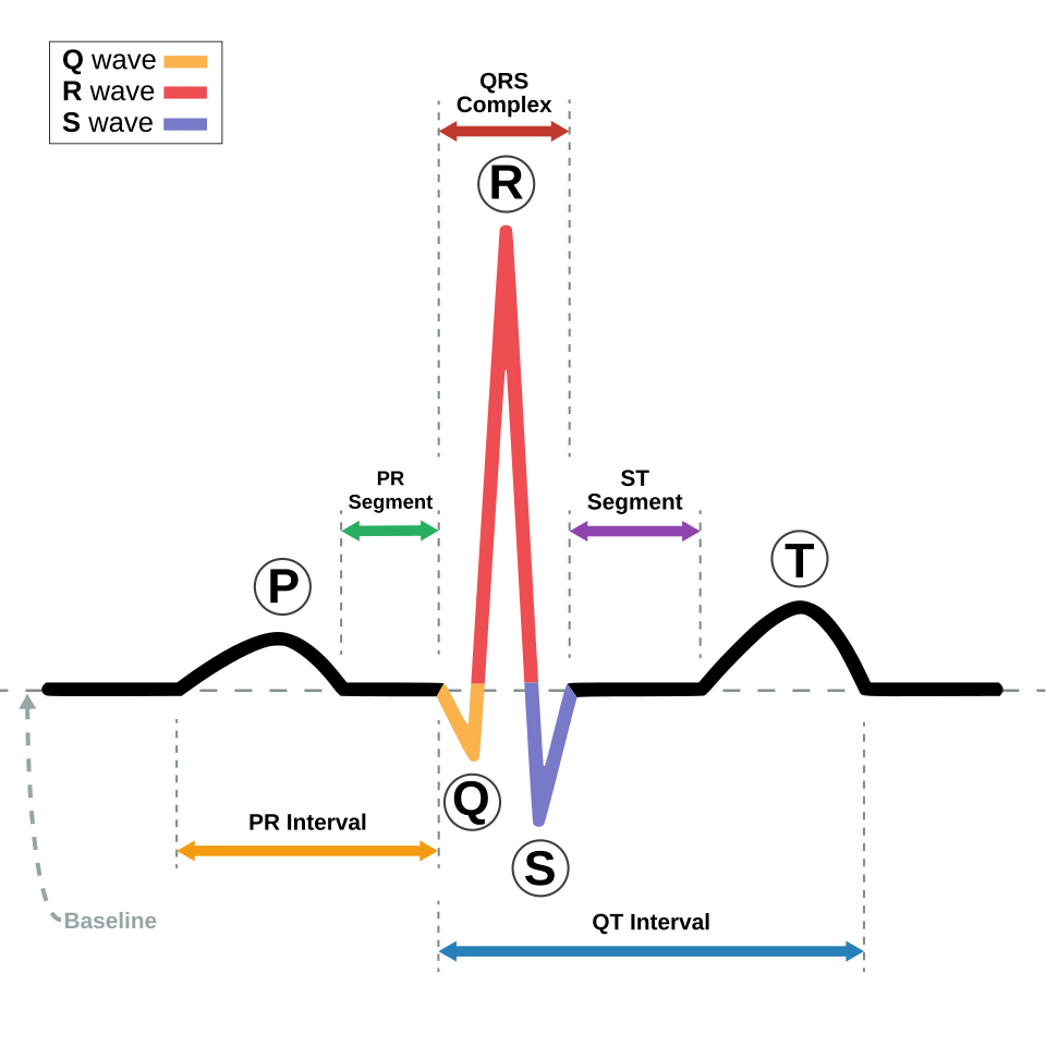
Normal cardiac conduction and the ECG
The sinoatrial (SA) node normally initiates each heartbeat. Depolarisation spreads through the atria, reaches the atrioventricular (AV) node, pauses briefly, then travels through the His bundle, bundle branches and Purkinje network to activate the ventricles.
The P wave represents atrial depolarisation. The PR interval runs from the beginning of atrial depolarisation to the beginning of ventricular depolarisation and therefore reflects atrial-to-ventricular conduction. The QRS complex represents ventricular depolarisation. A QRS duration of 120 ms or more is broad. The QT interval includes ventricular depolarisation and repolarisation; because it varies with heart rate, clinical interpretation usually relies on a corrected QT (QTc).
These components provide the vocabulary for the rest of rhythm interpretation. AV block is recognised by abnormal P-to-QRS conduction, pre-excitation by altered AV-to-ventricular activation, and many tachyarrhythmias by the combination of rate, regularity and QRS width.
Understand P waves, PR interval and QRS width before trying to name an arrhythmia.
A systematic rhythm approachA repeatable method for reading any rhythm strip.
When faced with an ECG or rhythm strip, use the same sequence every time:
- Pulse and stability: Is there a pulse? Is the rhythm causing hypotension, altered mental status, shock, ischaemic chest discomfort or acute heart failure?
- Rate: Is the ventricular rate fast, slow or roughly normal?
- Regularity: Is the rhythm regular, regularly irregular or irregularly irregular?
- QRS width: Is the QRS narrow or broad?
- Atrial activity: Are P waves or flutter waves present? Are P waves absent or hidden?
- Atrioventricular relationship: Does every P wave conduct? Is the PR interval fixed or changing? Are atria and ventricles dissociated?
This prevents premature pattern matching. For example, a regular narrow-complex tachycardia suggests a very different differential from an irregular broad-complex tachycardia, while AV dissociation during a broad-complex tachycardia strongly points toward ventricular tachycardia.
Stability → rate → regularity → QRS width → atrial activity → P:QRS relationship.
Life-threatening rhythms and instabilityInstability, shockable rhythms and broad-complex tachycardia.
The first distinction in an emergency is not the exact rhythm name; it is whether the patient is in cardiac arrest or has a pulse with haemodynamic compromise.
Unstable tachyarrhythmia and synchronised cardioversion
A tachyarrhythmia is considered clinically unstable when the tachycardia itself is causing poor perfusion—for example hypotension, altered mental status, signs of shock, ischaemic chest discomfort or acute heart failure. A patient may be tachycardic because of another unstable condition such as sepsis or haemorrhage, so the rhythm must be judged as the cause rather than merely an associated finding.
When an unstable patient has a pulse and a synchronisable tachyarrhythmia is responsible, prompt synchronised cardioversion is indicated. Synchronisation times the shock to ventricular depolarisation and reduces the risk of delivering energy during the vulnerable repolarisation period. Sustained polymorphic VT is an important exception because reliable synchronisation is not possible; it requires an unsynchronised high-energy shock.
Unstable tachyarrhythmia with a pulse → synchronised cardioversion, except polymorphic VT.
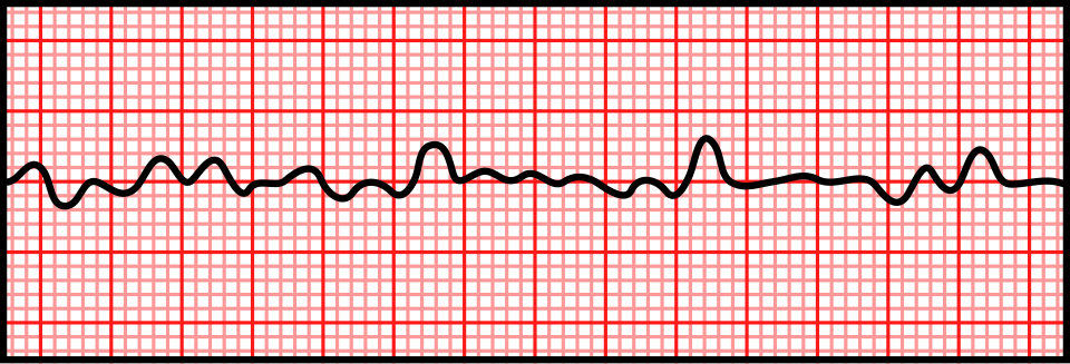
Ventricular fibrillation and pulseless VT are shockable
Ventricular fibrillation (VF) is chaotic ventricular electrical activity without coordinated mechanical contraction. Pulseless ventricular tachycardia (pVT) is an organised ventricular tachycardia that produces no palpable pulse. Both are cardiac-arrest rhythms.
Management centres on immediate high-quality CPR and rapid defibrillation. Defibrillation is unsynchronised because there is no useful organised ventricular cycle to synchronise to in VF, and the patient is pulseless in pVT.
VF and pulseless VT are the two shockable adult cardiac-arrest rhythms.
Asystole and PEA are non-shockable
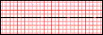
Asystole is the absence of meaningful ventricular electrical activity. Pulseless electrical activity (PEA) is organised or semi-organised electrical activity without a palpable pulse or effective circulation.
Neither is treated by defibrillation. Management instead centres on high-quality CPR, epinephrine according to the cardiac-arrest algorithm, and rapid identification and treatment of reversible causes. In suspected asystole, confirm that the tracing is genuine rather than a disconnected lead or very fine VF.
VF/pVT = shock; asystole/PEA = CPR + epinephrine + reversible causes.
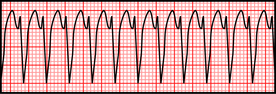
Broad-complex tachycardia should be treated as possible VT until clarified
A broad-complex tachycardia can be ventricular tachycardia, supraventricular tachycardia with aberrant conduction, or a pre-excited tachycardia. The consequences of incorrectly treating VT as a benign supraventricular rhythm can be serious.
When the diagnosis is uncertain, presume VT until a safer diagnosis is established, particularly in an older patient or one with structural/ischaemic heart disease. If the patient is stable, obtain a 12-lead ECG and use morphology, AV relationships and clinical context to refine the diagnosis. Adenosine has a limited diagnostic/therapeutic role only in selected stable, regular, monomorphic wide-complex tachycardias.
Wide + fast + uncertain → presume VT.
Ventricular tachycardia and torsades de pointesVT clues, torsades, magnesium and acquired QT prolongation.
Once a broad-complex tachycardia is recognised, the next task is to decide whether ventricular origin is supported and whether the QT interval changes management.
AV dissociation strongly supports ventricular tachycardia
During VT, the ventricles may be driven by a ventricular focus or re-entry circuit while the atria continue under sinus control. P waves therefore continue at their own rate and have no fixed relationship to the broad QRS complexes.
This AV dissociation is strong evidence for VT when it is seen during a broad-complex tachycardia. It is not always visible, so its absence does not exclude VT.
AV dissociation during a broad-complex tachycardia strongly favours VT.
Capture beats and fusion beats support VT
A capture beat occurs when an atrial impulse successfully conducts through the AV node during VT and completely activates the ventricles, producing a relatively normal or narrower QRS complex among the broad VT complexes.
A fusion beat occurs when the conducted sinus impulse and the ventricular tachycardia impulse activate the ventricles at approximately the same time. The resulting QRS has an intermediate morphology—part normal conducted beat and part ventricular beat.
Both findings demonstrate competing supraventricular and ventricular activation and strongly support VT in the appropriate context.
Capture beat = sinus activation takes over one beat; fusion beat = sinus and VT activation merge.
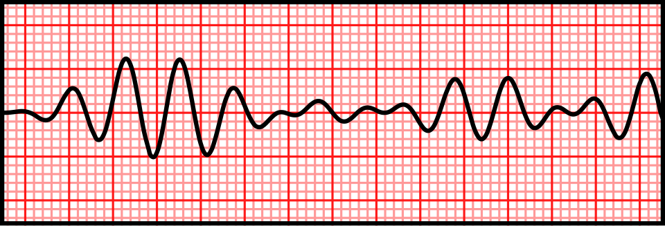
Torsades de pointes is polymorphic VT associated with prolonged QT
Torsades de pointes is a form of polymorphic ventricular tachycardia occurring in the setting of a prolonged QT interval. QRS morphology, amplitude and polarity vary beat to beat, producing the characteristic appearance of complexes twisting around the isoelectric baseline.
The QT interval that matters is the QT during the underlying rhythm, not the apparent interval during VT. Torsades may be self-terminating and recurrent, or it may become sustained, cause syncope, or deteriorate into VF. Sustained polymorphic VT is treated as electrically unstable and requires immediate unsynchronised shock.
Torsades = polymorphic VT + prolonged QT.
Magnesium is used for recurrent torsades even when serum magnesium is normal
Intravenous magnesium can suppress recurrence of torsades associated with long QT. Its use is not dependent on demonstrating hypomagnesaemia on a serum test, because the antiarrhythmic effect is not simply replacement of a measured deficiency.
This distinction matters: magnesium is used for torsades in the setting of long QT, but routine magnesium is not recommended for polymorphic VT with a normal QT interval. If sustained polymorphic VT is present, shock treatment takes priority over waiting for drug therapy.
Normal serum magnesium does not rule out IV magnesium for torsades.
Hypokalaemia, hypomagnesaemia and drugs can produce acquired long QT
Acquired QT prolongation is commonly promoted by QT-prolonging medications, hypokalaemia and hypomagnesaemia, often in combination. Bradycardia can further increase torsades risk in susceptible patients.
When acquired long QT is recognised, review and stop avoidable QT-prolonging drugs, correct electrolyte abnormalities and address bradycardia or other triggers where appropriate. The important clinical question is not merely whether the QT is long, but whether the patient has a substrate that can precipitate torsades.
Long QT + low K/Mg or QT-prolonging drugs = torsades risk.
Inherited long-QT syndromes and Brugada syndromeInherited long-QT syndromes and Brugada syndrome.
Inherited channelopathies are important because the myocardium may be structurally normal while electrical instability creates a risk of syncope, torsades, VF and sudden cardiac death.
Jervell and Lange-Nielsen syndrome
Jervell and Lange-Nielsen syndrome is a rare congenital long-QT syndrome usually caused by biallelic pathogenic variants in KCNQ1 or KCNE1. It is inherited in an autosomal-recessive pattern and is associated with congenital, typically severe sensorineural hearing loss.
The classic examination association is therefore prolonged QT plus congenital deafness. The arrhythmic phenotype can be severe, with syncope and life-threatening ventricular arrhythmias occurring early in life.
Long QT + congenital sensorineural deafness → Jervell and Lange-Nielsen syndrome.
Romano-Ward syndrome
Romano-Ward syndrome refers to the more common autosomal-dominant forms of congenital long-QT syndrome without congenital deafness. Depending on the affected ion channel and individual risk profile, patients may be asymptomatic or may develop exertional/emotional syncope, torsades or sudden cardiac death.
The distinction from Jervell and Lange-Nielsen syndrome is primarily the inheritance pattern and absence of congenital hearing loss.
Romano-Ward = autosomal dominant long QT without congenital deafness.
Type 1 Brugada ECG pattern
The diagnostic type 1 Brugada pattern shows coved ST-segment elevation in the right precordial leads, followed by a negative T wave. The pattern is classically sought in V1 and V2, including higher right-precordial positions when clinically appropriate.
The ECG pattern must be interpreted in clinical context. Brugada syndrome is an inherited arrhythmia syndrome associated with ventricular fibrillation and sudden cardiac death, but not every right-precordial ST abnormality represents Brugada syndrome.
Type 1 Brugada = coved right-precordial ST elevation followed by a negative T wave.
Fever can unmask Brugada syndrome
Fever can worsen the sodium-channel dysfunction associated with Brugada syndrome. A diagnostic ECG pattern may become more obvious during fever, and susceptible patients may have increased risk of malignant ventricular arrhythmias.
Fever in a patient with known or suspected Brugada syndrome therefore deserves prompt antipyretic treatment, ECG assessment and appropriate clinical monitoring based on symptoms and risk.
Fever can reveal a Brugada pattern and increase arrhythmic risk.
ICD therapy is for high-risk Brugada syndrome, not every Brugada pattern
An implantable cardioverter-defibrillator (ICD) is the main proven protection against recurrent lethal ventricular arrhythmias in high-risk Brugada syndrome, particularly after cardiac arrest or documented sustained ventricular arrhythmia. Suspected arrhythmic syncope in a patient with a spontaneous type 1 pattern also requires specialist risk assessment.
An asymptomatic patient with only a drug-induced pattern does not automatically require an ICD. Device therapy has complications, so risk stratification matters.
Brugada + prior cardiac arrest/documented malignant ventricular arrhythmia = strong ICD territory.
Atrial fibrillation and atrial flutterAF, flutter, stroke prevention, rate control and ablation.
Atrial fibrillation and flutter are both atrial tachyarrhythmias, but their atrial organisation is very different. AF is chaotic; typical flutter is organised macro-re-entry.
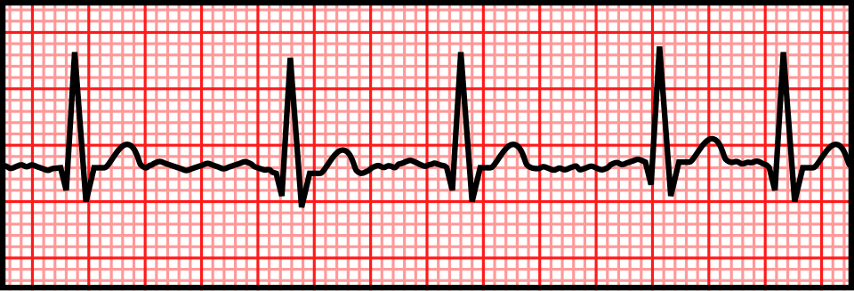
Atrial fibrillation: absent consistent P waves and an irregularly irregular ventricular rhythm
In atrial fibrillation, atrial activation is disorganised and there is no single repetitive atrial depolarisation. Consistent P waves therefore disappear. The AV node receives irregular atrial impulses and conducts a variable number to the ventricles, producing an irregularly irregular ventricular rhythm.
When the ventricular response is very slow or highly regular, consider additional AV-node disease, medication effect or pacing rather than assuming the usual AF pattern.
AF = no consistent P waves + irregularly irregular R–R intervals.
AF promotes thrombus formation in the left atrial appendage
Loss of coordinated atrial contraction promotes blood stasis, especially in the left atrial appendage. Stasis, together with atrial structural and endothelial changes, contributes to thrombus formation. Embolisation of left-atrial thrombus can cause ischaemic stroke or systemic embolism.
This is why stroke prevention is a core part of AF management and is considered separately from symptom control.
AF-related thrombus commonly forms in the left atrial appendage.
Stroke risk is not determined by symptom frequency
A patient who rarely notices AF is not necessarily at low thromboembolic risk. Conversely, frequent palpitations do not by themselves mean anticoagulation is required. Stroke-prevention decisions are based on validated clinical thromboembolic risk assessment and the patient's underlying risk factors.
Paroxysmal AF can still carry clinically important stroke risk. Symptom burden and stroke risk are different questions.
Anticoagulation decisions follow thromboembolic risk, not how often AF is felt.
Aspirin is not an adequate substitute for indicated anticoagulation
Antiplatelet therapy does not provide equivalent protection against AF-related thromboembolism. When a patient with AF meets criteria for oral anticoagulation and has no separate reason to use antiplatelet therapy, aspirin should not be substituted for appropriate anticoagulation simply because it is perceived as “safer” or easier.
Choice of anticoagulant depends on valve status, renal function, bleeding risk, interactions and other patient factors.
If anticoagulation is indicated for AF, aspirin is not an equivalent alternative.
Why AF duration matters before cardioversion
Cardioversion can restore organised atrial electrical and mechanical activity in a patient who may already have formed atrial thrombus. This creates a risk of embolisation around the time of cardioversion.
The traditional examination rule uses 48 hours or unknown duration as an important threshold for anticoagulation/imaging precautions before elective cardioversion. Contemporary guidance is more nuanced: the 2024 ESC guideline takes a more conservative safety-first approach once AF duration exceeds 24 hours, while other major guidance retains different timing frameworks. The clinically durable principle is to consider episode duration, anticoagulation status, thromboembolic risk and imaging before planned cardioversion rather than treating one time cut-off as universal.
Before elective AF cardioversion, think duration + anticoagulation + thromboembolic risk, not just the rhythm strip.
Beta-blockers commonly provide ventricular rate control in AF
A rate-control strategy accepts ongoing AF but limits the ventricular response. Beta-blockers reduce sympathetic drive and slow AV nodal conduction, so they are commonly used for ventricular rate control.
Choice of rate-control therapy depends on blood pressure, ventricular function, associated heart failure, comorbidities and whether management is acute or chronic. Nondihydropyridine calcium-channel blockers are also options in appropriate patients, while pre-excited AF is a separate emergency in which AV nodal blockers can be harmful.
Rate control slows AV nodal conduction; it does not necessarily restore sinus rhythm.
Digoxin controls resting rate better than exercise rate
Digoxin slows AV nodal conduction largely by increasing vagal influence. It can therefore be useful for ventricular rate control, especially at rest. During exercise or other adrenergic states, sympathetic activity rises and vagal influence falls, making digoxin less effective as a sole agent for controlling the ventricular response.
Its role depends on the clinical setting and it may be combined with other rate-control therapy when appropriate.
Digoxin is more effective for resting than exertional ventricular rate control.
Atrial flutter produces organised flutter waves
Typical atrial flutter is usually a macro-re-entrant rhythm travelling around the right atrium. Atrial activation is rapid but organised, producing repetitive flutter waves. In the classic form, these can create a sawtooth appearance, particularly in the inferior leads.
This contrasts with AF, where atrial activation is chaotic and there are no repetitive organised P or flutter waves.
Flutter = organised repetitive atrial activity; AF = chaotic atrial activity.
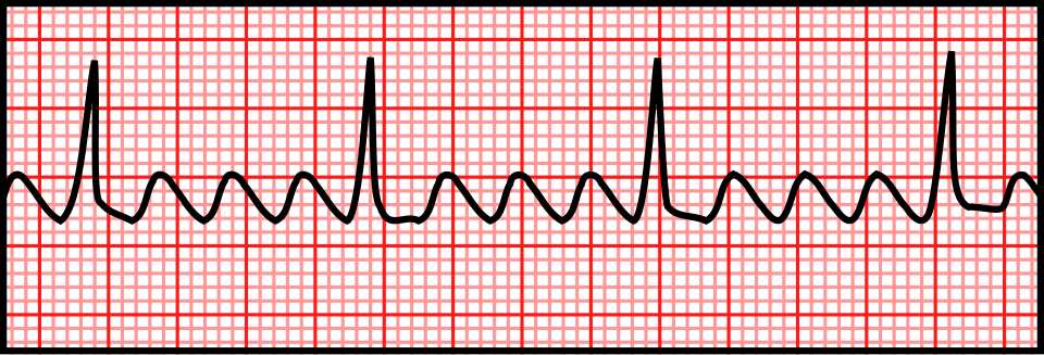
2:1 atrial flutter often produces a ventricular rate near 150/min
Typical flutter commonly has an atrial rate near 300/min. If every second flutter wave conducts through the AV node, the ventricular rate is therefore close to 150/min.
A regular narrow-complex tachycardia around 150/min should prompt a deliberate search for flutter waves, because 2:1 conduction can hide one flutter wave within the QRS or T wave.
Regular narrow tachycardia near 150/min → actively look for 2:1 atrial flutter.
Typical atrial flutter is highly amenable to catheter ablation
Typical atrial flutter depends on a predictable circuit through the cavotricuspid isthmus. Radiofrequency catheter ablation can interrupt this critical part of the circuit and has a high success rate for recurrent or symptomatic typical flutter.
The simplicity of the circuit is why typical flutter is generally more straightforward to ablate than atrial fibrillation.
Typical flutter has a defined isthmus-dependent circuit that can be ablated.
Regular narrow-complex supraventricular tachycardiaAVNRT, AVRT, vagal manoeuvres and adenosine.
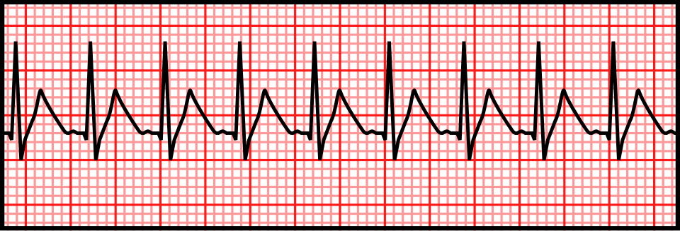
“SVT” is a broad term. In exam questions, a sudden-onset regular narrow-complex tachycardia often refers to AVNRT or orthodromic AVRT, both of which usually depend on the AV node as part of the re-entry circuit.
Regular narrow-complex SVT commonly represents AVNRT or AVRT
AV nodal re-entrant tachycardia (AVNRT) uses dual pathways within or close to the AV node. Orthodromic AV re-entrant tachycardia (AVRT) travels antegradely through the AV node and returns to the atria via an accessory pathway.
In both, ventricular activation usually proceeds through the normal His–Purkinje system, so the QRS remains narrow unless there is pre-existing or rate-related aberrancy.
Sudden regular narrow-complex tachycardia → think AVNRT or orthodromic AVRT.
Vagal manoeuvres are first-line for stable regular narrow-complex tachycardia
Vagal manoeuvres transiently increase vagal tone and slow conduction through the AV node. If the AV node is essential to a re-entry circuit, this temporary slowing may interrupt the circuit and terminate the tachycardia.
For a haemodynamically stable regular narrow-complex tachycardia, vagal manoeuvres are recommended before drug therapy. A modified Valsalva manoeuvre is more effective than a simple Valsalva in many patients.
Stable regular narrow-complex tachycardia → vagal manoeuvre first.
Adenosine transiently blocks the AV node
Adenosine has an extremely short duration of action and produces a brief AV nodal block. It can terminate AVNRT and orthodromic AVRT because those circuits require AV nodal conduction. It can also transiently expose underlying atrial activity, helping reveal flutter or another atrial rhythm even when it does not terminate that rhythm.
Because its half-life is only seconds, adenosine must be delivered as a rapid IV bolus followed immediately by a flush.
Adenosine terminates AV-node-dependent re-entry by briefly blocking the AV node.
Adenosine and asthma/bronchospasm
Adenosine can provoke clinically significant bronchospasm. Current AHA advanced life-support guidance describes asthma as a contraindication because severe bronchospasm can occur.
In a patient with active bronchospasm or significant reactive airway disease, do not give adenosine reflexively. Reassess the rhythm, stability and alternative treatment options.
Adenosine can cause severe bronchospasm—avoid it in asthma/active bronchospasm.
Wolff–Parkinson–White syndrome and pre-excitationAccessory pathways, delta waves and pre-excited AF.
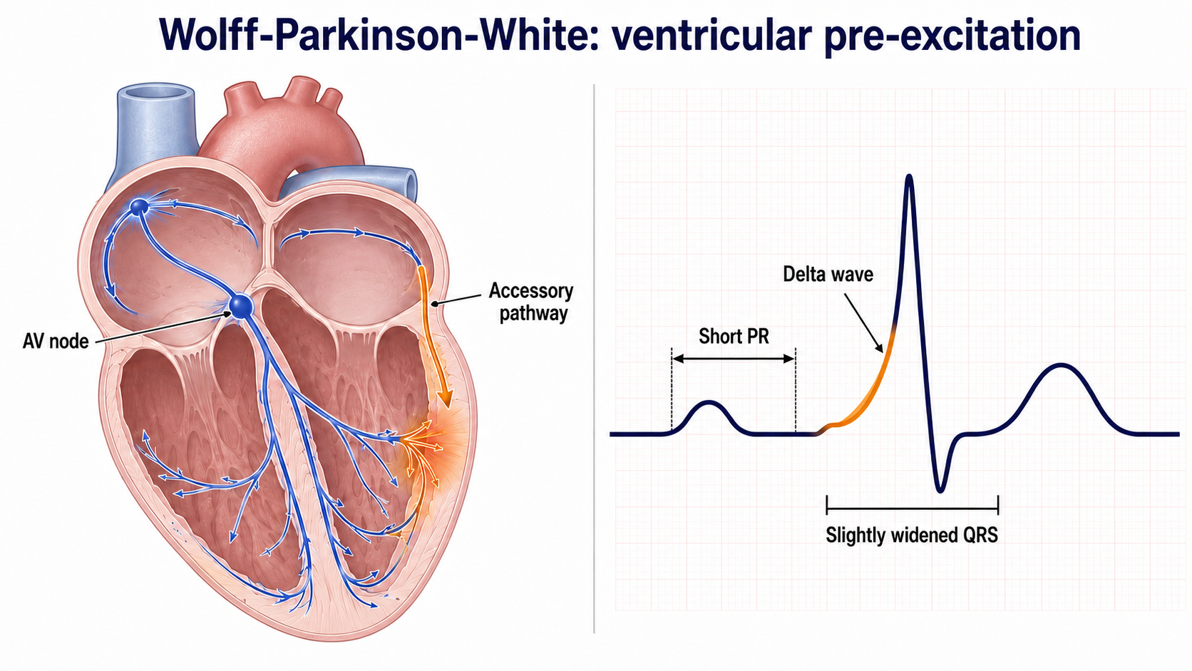
An accessory pathway creates an electrical route between atrium and ventricle that bypasses the normal AV nodal delay. This can produce both ventricular pre-excitation in sinus rhythm and re-entrant tachycardias.
WPW: short PR interval and delta wave
When an accessory pathway conducts from atrium to ventricle during sinus rhythm, part of the ventricle depolarises earlier than it would through the normal AV node–His–Purkinje pathway. The PR interval is therefore shortened and the beginning of the QRS is slurred, producing the delta wave. The QRS may be widened because ventricular activation begins partly through myocardium rather than entirely through the specialised conduction system.
Strictly, WPW pattern refers to pre-excitation on the ECG; WPW syndrome generally implies pre-excitation associated with symptomatic tachyarrhythmia.
Short PR + delta wave = ventricular pre-excitation through an accessory pathway.
AF with pre-excitation: AV nodal blockers can be dangerous
During AF, the AV node normally limits how many atrial impulses reach the ventricles. An accessory pathway may lack this protective filtering and can conduct atrial impulses extremely rapidly.
In pre-excited AF, AV nodal blocking drugs such as beta-blockers, verapamil/diltiazem and digoxin can favour conduction through the accessory pathway and may accelerate the ventricular response; AHA guidance also advises against IV amiodarone in this setting. The ECG is often very fast, irregular and broad-complex. Urgent expert management is required, with electrical cardioversion if unstable.
Irregular + very fast + broad + pre-excitation → do not give ordinary AV nodal blockers.
Catheter ablation can definitively treat an accessory pathway
Catheter ablation maps the accessory pathway and destroys a small region of tissue required for abnormal conduction. Successful ablation removes the substrate for accessory-pathway-mediated AVRT and, when the pathway conducts antegradely, eliminates ventricular pre-excitation.
It is therefore a definitive treatment for many patients with symptomatic accessory-pathway-mediated arrhythmias rather than merely suppressing individual episodes.
Ablation removes the accessory pathway—the substrate of the arrhythmia.
Bradycardia and AV blockFirst-degree block through complete heart block and pacing.
AV block is easiest to understand as a failure of atrial impulses to reach the ventricles normally. The key ECG questions are whether every P wave conducts and whether the PR interval changes before a dropped beat.
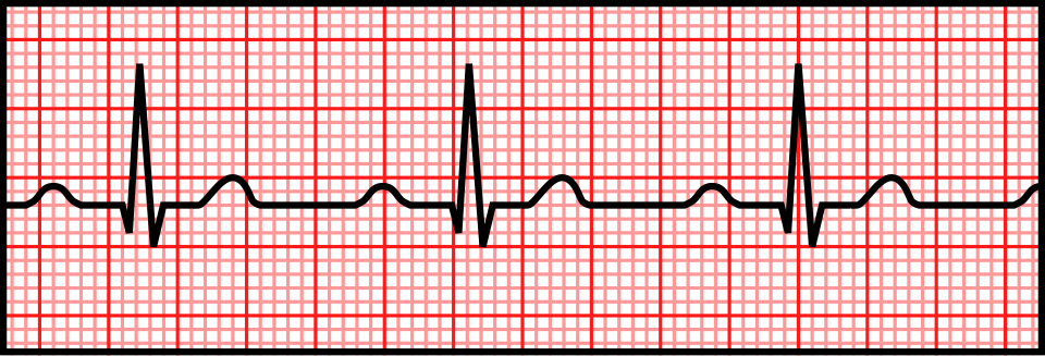
First-degree AV block
In first-degree AV block, every atrial impulse still conducts to the ventricles, but conduction is delayed. The ECG shows a PR interval >200 ms with a QRS after every P wave.
No beats are actually dropped, so “first-degree AV block” is more accurately a prolonged AV conduction time than intermittent failure of conduction.
First-degree AV block = PR >200 ms, every P conducts.
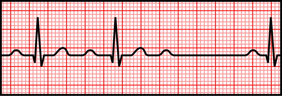
Mobitz I (Wenckebach)
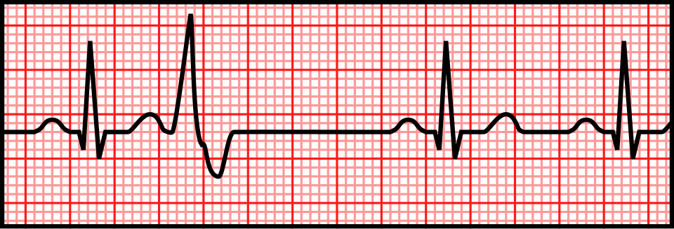
In Mobitz I, AV conduction progressively slows across successive beats. The PR interval therefore becomes progressively longer until one P wave is not followed by a QRS complex. After the dropped beat, the sequence resets.
Mobitz I most often reflects AV nodal block and can occur with increased vagal tone or AV nodal blocking drugs, although clinical context matters.
Mobitz I = progressively longer PR intervals → dropped QRS.
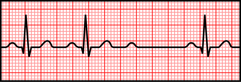
Mobitz II
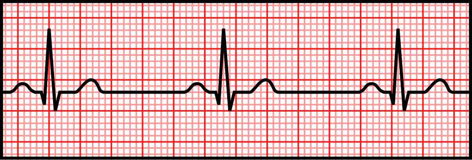
In Mobitz II, the PR interval of conducted beats is relatively constant, but an atrial impulse suddenly fails to conduct and a QRS complex is dropped. There is no progressive PR prolongation beforehand.
Mobitz II usually reflects disease within the His–Purkinje system and is more likely than Mobitz I to progress to advanced block. A 2:1 AV block cannot always be confidently labelled Mobitz I or Mobitz II from PR behaviour alone because only one PR interval is visible between dropped beats.
Mobitz II = fixed PR on conducted beats + sudden non-conducted P wave.
Mobitz II can progress to complete heart block
Because Mobitz II often represents disease below the AV node, conduction can fail more extensively without a preceding Wenckebach pattern. Progression to high-grade or complete AV block can cause profound bradycardia, syncope or haemodynamic compromise.
This is why Mobitz II warrants urgent assessment, correction of reversible causes and pacing evaluation even when the patient is temporarily stable.
Mobitz II is high risk because it can progress unpredictably to complete AV block.
Complete heart block causes AV dissociation
In third-degree AV block, no atrial impulses conduct to the ventricles. The atria continue under sinus control while a junctional or ventricular escape pacemaker drives the ventricles independently.
The ECG therefore shows P waves marching through at one rate and QRS complexes at a slower independent rate, with no fixed PR relationship. The QRS may be narrow with a junctional escape or broad with a lower ventricular escape.
Complete heart block = atria and ventricles beat independently.
 First-degree AV block
 Mobitz I (Wenckebach)Progressive PR prolongation, then a dropped QRS.2nd Degree AV Block (Mobitz I).svg · CC0
 Mobitz IIFixed PR intervals with sudden failure of conduction.2nd Degree AV Block (Mobitz II).svg · CC0
 Complete heart block


.svg)


.svg)
.svg)

Symptomatic bradycardia: treat compromise and correct the cause
Bradycardia is clinically important when it causes poor perfusion—such as hypotension, altered mental status, shock, ischaemic chest discomfort, acute heart failure or syncope. At the same time, look for reversible causes including ischaemia, hypoxia, electrolyte/metabolic disturbance, medication toxicity and hypothermia.
Current AHA guidance supports atropine for acute bradycardia with haemodynamic compromise. If atropine is ineffective, transcutaneous pacing and/or rate-accelerating infusions such as epinephrine or dopamine can be used while preparing more definitive therapy when necessary.
Treat symptomatic/unstable bradycardia, not an isolated slow heart rate.

Mobitz II and complete heart block commonly require pacing
Persistent Mobitz II, high-grade AV block and third-degree AV block can make ventricular activation unreliable. If the block is not due to a reversible cause, permanent pacing is commonly indicated because the ventricles should not depend on an unstable diseased conduction system.
The urgency depends on symptoms, escape rhythm, haemodynamic status and the cause of the block; temporary pacing may be needed while definitive management is arranged.
Advanced infranodal AV block often requires pacing.
Sick sinus syndrome and tachy-brady syndrome
Sinus-node dysfunction may cause inappropriate sinus bradycardia, sinus pauses, sinoatrial exit block or chronotropic incompetence. Some patients also develop atrial tachyarrhythmias such as AF or flutter.
When periods of atrial tachyarrhythmia alternate with sinus bradycardia or pauses, this is called tachy-brady syndrome. Symptoms, correlation with documented bradycardia and reversible causes guide pacing decisions.
Tachy-brady syndrome = sinus-node dysfunction with alternating bradycardia and atrial tachyarrhythmias.
Ectopy, pacing and secondary preventionPVC patterns, pacemaker capture and secondary prevention.
These final patterns apply the same principles: identify where the beat originates, whether an electrical stimulus produces myocardial depolarisation, and whether a malignant rhythm has a reversible cause.
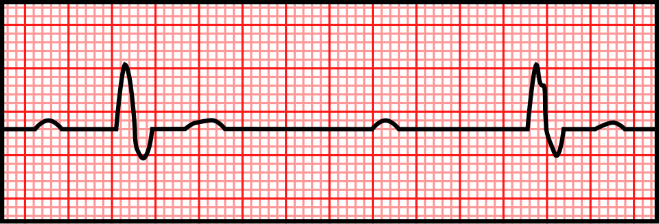
Ventricular bigeminy
Bigeminy describes a repeating pattern in which every normal beat is followed by a premature beat. In ventricular bigeminy, each sinus beat is followed by a premature ventricular complex (PVC), producing a repeating 1:1 sequence.
The term describes the pattern, not the underlying cause. PVCs may occur in structurally normal hearts or with ischaemia, cardiomyopathy, electrolyte disturbance, stimulants and other conditions.
Ventricular bigeminy = sinus beat → PVC → sinus beat → PVC.

Pacemaker failure to capture
A pacemaker spike shows that the device delivered an electrical stimulus. Successful capture means that stimulus is followed by depolarisation of the paced chamber—a P wave after an atrial pacing spike or a QRS complex after a ventricular pacing spike.
Failure to capture occurs when a pacing spike is not followed by the expected depolarisation. Causes include lead displacement or fracture, inadequate output, rising pacing threshold, myocardial ischaemia and electrolyte/metabolic abnormalities. In a pacemaker-dependent patient, loss of capture can be immediately dangerous.
Pacing spike with no subsequent depolarisation = failure to capture.
Survivors of unexplained VF need secondary-prevention evaluation
Survival from VF should trigger a systematic search for reversible causes such as acute ischaemia, major electrolyte abnormality, drug toxicity or another transient trigger. If no completely reversible cause is found, investigate for structural heart disease and inherited electrical disorders.
Patients who survive VF or haemodynamically unstable sustained VT without a reversible cause are often candidates for secondary-prevention ICD therapy, because the risk of recurrent malignant ventricular arrhythmia remains substantial.
Unexplained VF survivor → find the cause and assess for secondary-prevention ICD.
Clinical synthesisBring the main rhythm patterns together.
The fastest way to interpret arrhythmias is to keep returning to the same framework. First decide whether the patient has a pulse and whether the rhythm is compromising perfusion. Then classify the rhythm by rate, regularity and QRS width. Finally, examine atrial activity and the P-to-QRS relationship.
That framework explains the major patterns:
- VF/pVT are shockable arrest rhythms; asystole/PEA are not.
- A broad regular tachycardia of uncertain origin should be treated as possible VT.
- AV dissociation, capture beats and fusion beats support VT.
- Polymorphic VT + prolonged QT is torsades de pointes.
- No consistent P waves + irregularly irregular rhythm suggests AF.
- Sawtooth atrial activity suggests flutter; a ventricular rate near 150/min should raise suspicion for 2:1 flutter.
- Regular narrow-complex tachycardia often represents AVNRT or orthodromic AVRT.
- Short PR + delta wave indicates ventricular pre-excitation.
- Progressive PR prolongation then a dropped beat is Mobitz I; fixed PR with sudden dropped conduction is Mobitz II.
- Independent atrial and ventricular rhythms at a slow ventricular rate indicate complete AV block.
The aim is not simply to identify ECG shapes, but to understand the electrical mechanism, the immediate danger and why the treatment works.
References & image provenanceGuidelines and media licensing used for this article.
The treatment-sensitive sections were checked against the following current/authoritative sources:
- American Heart Association. 2025 Guidelines for CPR and Emergency Cardiovascular Care — Part 9: Adult Advanced Life Support.
- ACC/AHA/ACCP/HRS. 2023 Guideline for the Diagnosis and Management of Atrial Fibrillation.
- European Society of Cardiology. 2024 Guidelines for the Management of Atrial Fibrillation.
- European Society of Cardiology. 2019 Guidelines for Supraventricular Tachycardia.
- European Society of Cardiology. 2022 Guidelines for Ventricular Arrhythmias and Prevention of Sudden Cardiac Death.
- European Society of Cardiology. 2021 Guidelines on Cardiac Pacing and Cardiac Resynchronization Therapy.
- GeneReviews. Long QT Syndrome Overview; Jervell and Lange-Nielsen Syndrome; Brugada Syndrome.
THIRD_PARTY_MEDIA.md.